DICOM Viewer Control
The LEADTOOLS DICOM Viewer Control is a high-level component for developers to build medical display solutions such as view stations, teleradiology applications, DICOM DVD viewers, and high-end diagnostic workstations found in radiology and other medical specialty departments. Whether you are building a PACS viewer for radiology, cardiology, mammography, ophthalmology, pathology, dermatology, dentistry, or any other specialty, the DICOM Viewer Control eliminates complexity from your .NET Framework, C++ Class Library, C#, VB, C/C++, and HTML / JavaScript projects and sets a solid DICOM and imaging foundation on which you can rely.

Built-in DICOM Viewer Overlay Templates & Tools
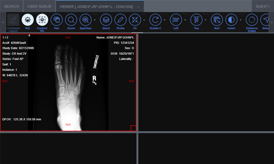
The control includes a set of built-in overlay templates to display information such as window center/width values, patient information, and field of view.
- Information image overlay is an essential feature for any high-end medical viewer. As a programmer, you have full control of the text, size, style, color, and location within the window. Additionally, overlays may be permanently rendered onto the image or left as an overlay.
-
Tools for:
- Annotation and mark-up (text, arrow, rectangle, ellipse, Cobb angle, Norberg, custom annotation object)
- Image alpha (sigmoid, exponential, logarithmic)
- Image display (window level, zoom, zoom to rectangle, pan, magnifying glass, spatial locator, user spy glass)
- Measurement (ruler, poly-ruler, snap ruler, protractor, statistics, probe)
- Region of interest (magic wand, nudge, shrink)
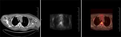
- Specialized diagnostic evaluation tools such as PET-CT fusion
- Cine playback
- Image processing functions include rotate, flip, reverse, and invert

Fully Customizable Cells and Display
Fully customizable cell and sub-cell layout that includes:
- Adjacent cells
- Any cell location
- Any cell size
- Empty cells
- Overlapping cells
- Automated image synchronization to view multiple series
- Customizable image display options to control the image view and scale
- Various interpolated image display algorithms to adjust display quality and speed
- Options for low-memory usage facilitate large study display
DICOM and Medical Image Annotation Libraries
A broad range of DICOM and medical image annotation objects are included in the Medical Image Viewer and Medical Workstation Viewer controls. These medical image annotations can be added to DICOM, bitonal, color, and grayscale images, either on a presentation layer or burned into the image data. They are perfect for highlighting areas of interest and redacting private information. Access to edit annotations or view private information can be control with user-level passwords. Flexible annotation object storage options include DICOM data sets (Grayscale Softcopy Presentation State), image files, separate annotation files, database, memory, and XML.
- Audio
- Video
- Cross Product
- Encryption
- Freehand
- Pointer
- Polyruler
- Protractor
- Redaction
- Ruler
- Text
- Text Pointer

Integrates with Medical 3D Volume Rendering Libraries
LEADTOOLS Medical 3D Volume Rendering libraries offer developers many different data representations using various 3D reconstruction techniques.
- Multiple volume types including VRT, MIP, MPR, SSD
- Interactive controls for window level, density removal, offset, scale, zoom, and more
- Settings and options for customizing the volume appearance including border, rotation cube, resolution, and slicing
- Caches 3D volumes for faster load times
- Display 3D reference lines on any series, including optional simultaneous display of boundary lines (first and last)
- 3D cursor for smooth MPR and synchronized stack navigation

Other DICOM Viewers
The LEADTOOLS libraries feature two other DICOM/Medical viewers:

Easy to Integrate
LEADTOOLS handles the heavy lifting, eliminating months of R&D, while giving you the best quality and performance available. You'll be free to focus on other components of your application. Download the LEADTOOLS evaluation to streamline your development.

DICOM Viewer Control SDK Platforms and Programming Interfaces
Operating Systems
Projects that use LEADTOOLS DICOM Viewer Control libraries can be deployed to Windows and Web devices.
Frameworks
Developers that are leveraging these frameworks can utilize the DICOM Viewer Control SDK: .NET Framework, WinForms, C++ Class Library, ASP.NET, and Web Services / Web API (JSON, SOAP, REST)
Programming, Scripting, Markup
DICOM Viewer Control code snippets and demo applications are provided for the following: C#, VB, XAML, C/C++, and HTML / JavaScript
Start Coding with LEADTOOLS DICOM Viewer Control
DICOM Viewer Control libraries as well as all LEADTOOLS Recognition, Document, Medical, Vector, and Imaging technologies for all development and target platforms, including Windows, Linux, and macOS.
Online Demo Application that include DICOM Viewer Control
HTML5/JavaScript Medical Web Viewer Framework
A zero-footprint Medical viewer with light and dark themes to display DICOM images (pixel data) with window level, density removal, offset, scale, zoom, stack, annotation/markup, and multi-touch support for phone, tablet, and desktop.
It demonstrates zero-footprint 3D volume rendering of multiple volume types including VRT, MIP, MPR, and SSD in the Medical Web Viewer. The viewer does not require browser plugins, desktop utilities, or remote desktop clients and options for low-resolution and caching to speed up rendering and loading.
Note: If you have your own test images that you would like to upload into the application, contact support@leadtools.com to have a private user profile created.